2 Planning an fMRI study
In this section: experimenter’s decisions
Whole-brain or ROI analysis (or both)
Univariate or multivariate analysis
Blocked or event-related design
Analysis steps
Single-subject - first level - fixed effects analysis (FFX)
Group - second-level - random effects analysis (RFX)
(also mixed effects analysis is possible, but relatively rare due to large matrix size)
This is equivalent to e.g. conducting many trials per subject to measure reaction time, and then compute a subject-specific mean per condition, after which you would perform the actual statistical inference
Multilevel modelling is also possible (for small datasets), but less frequent.
No matter what you do, your analysis will boil down to these stages.
Research questions
Whole-brain or ROI analysis?
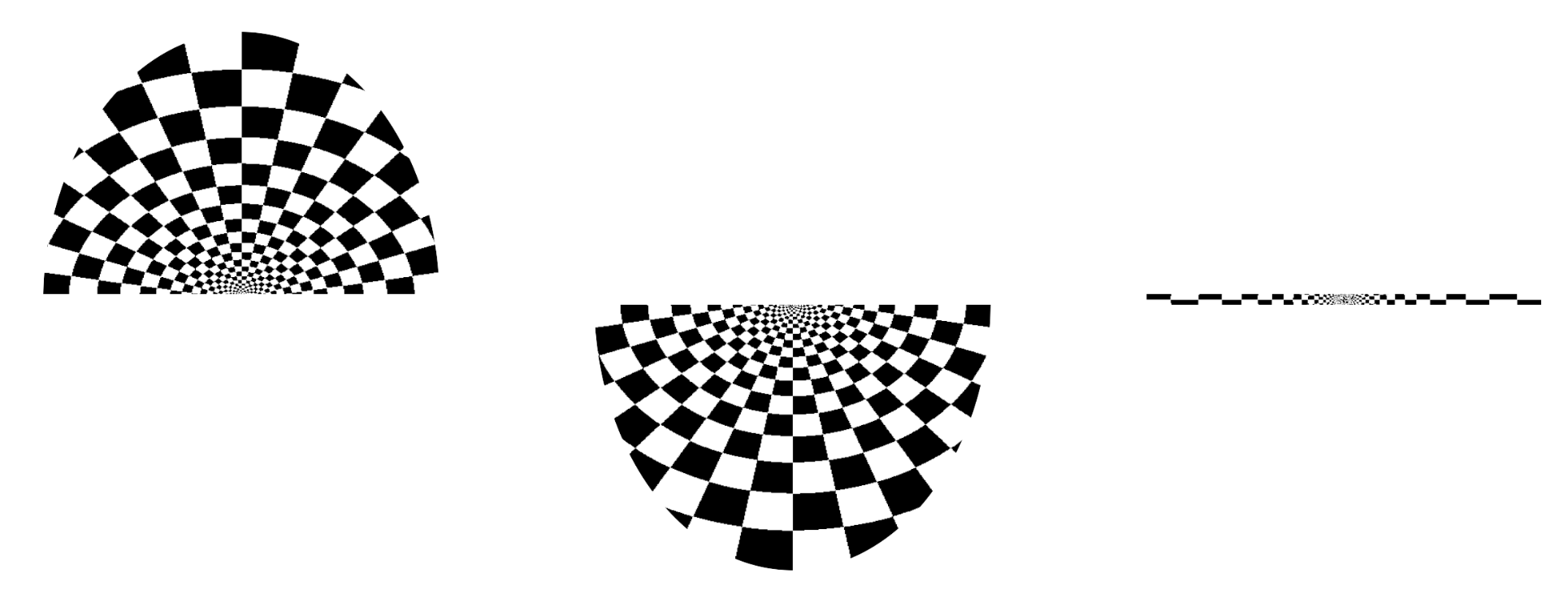
Exploratory: Neural correlates of illusory shapes
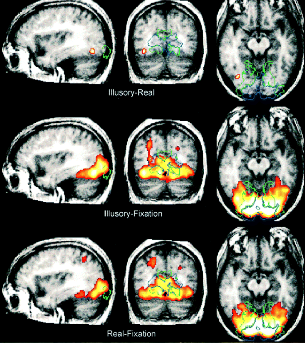
Hypothesis-driven: Are illusory shapes represented in the dorsal visual stream?
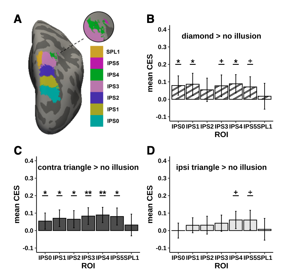
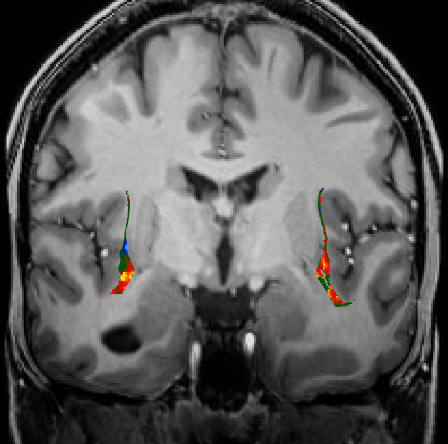
Defining ROIs
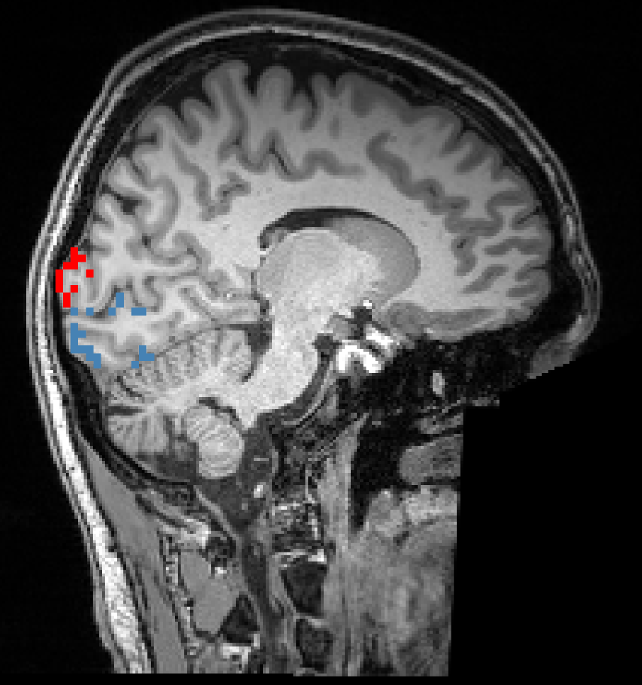
Functional localizer scan
Anatomical scan segmentation
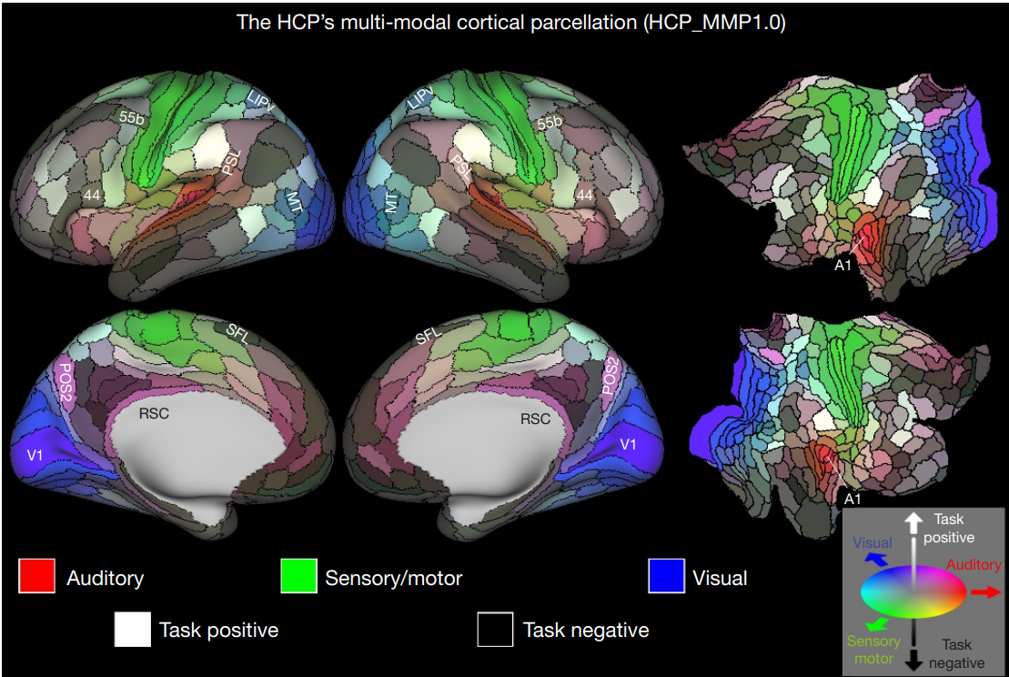
Atlas in standard space
Functional localizer scan
Cortical atlas
ROI analysis is a way to deal with multiple comparison problem. But it requires an a priory and independent definition of an ROI.
There are two ways to define ROIs:
From an anatomical scan
From an additional (separate) functional experiment
Subcortical ROIs
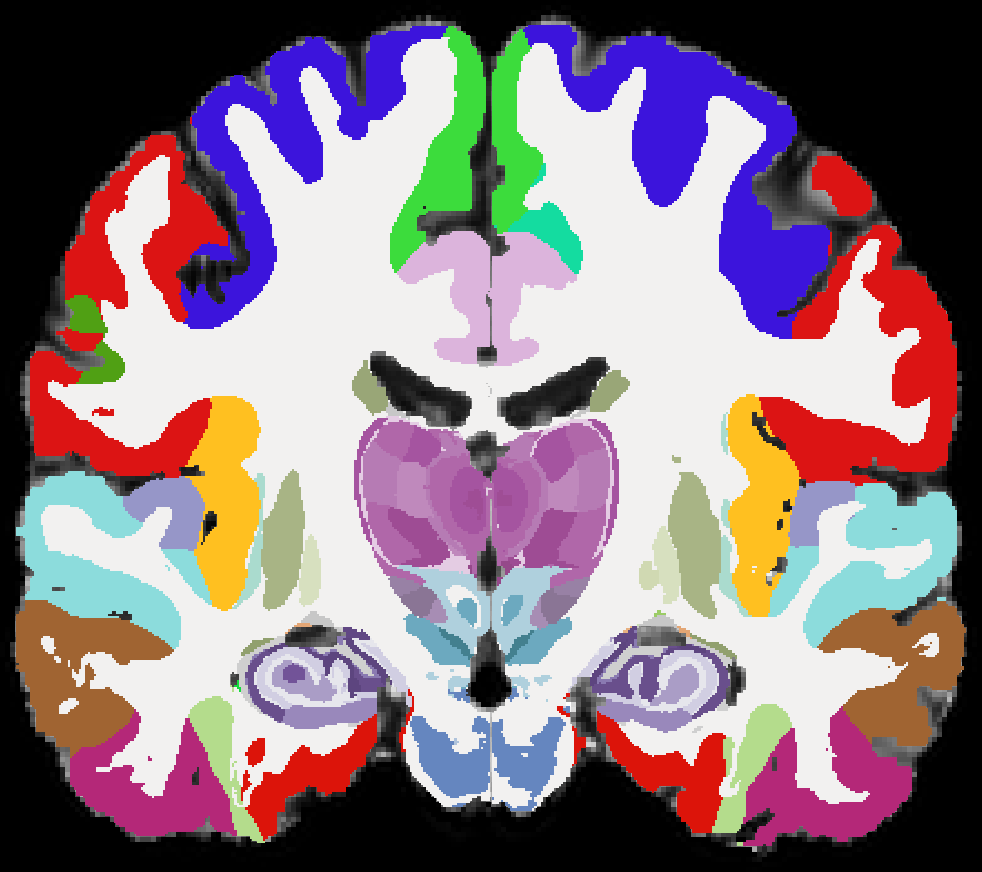
This is the state-of-the-art for anatomically-based ROI definition based on deep learning
Univariate or mutlivariate?
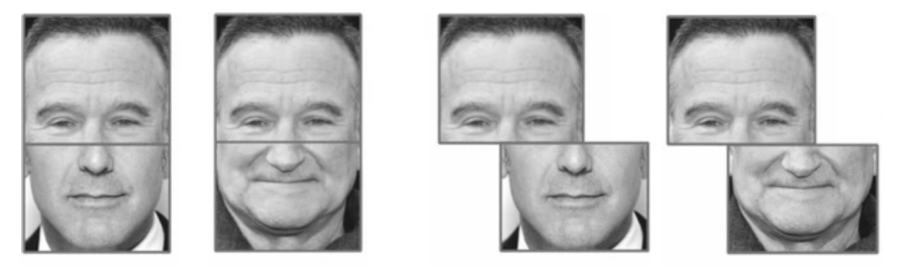
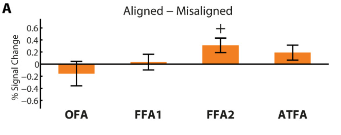
Univariate: Does FFA respond to composite face illusion?
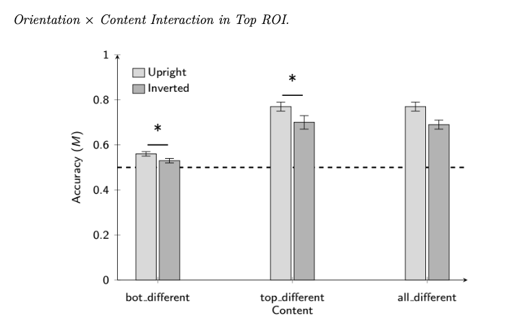
Multivariate: Is early visual cortex representation reflects the composite face illusion?
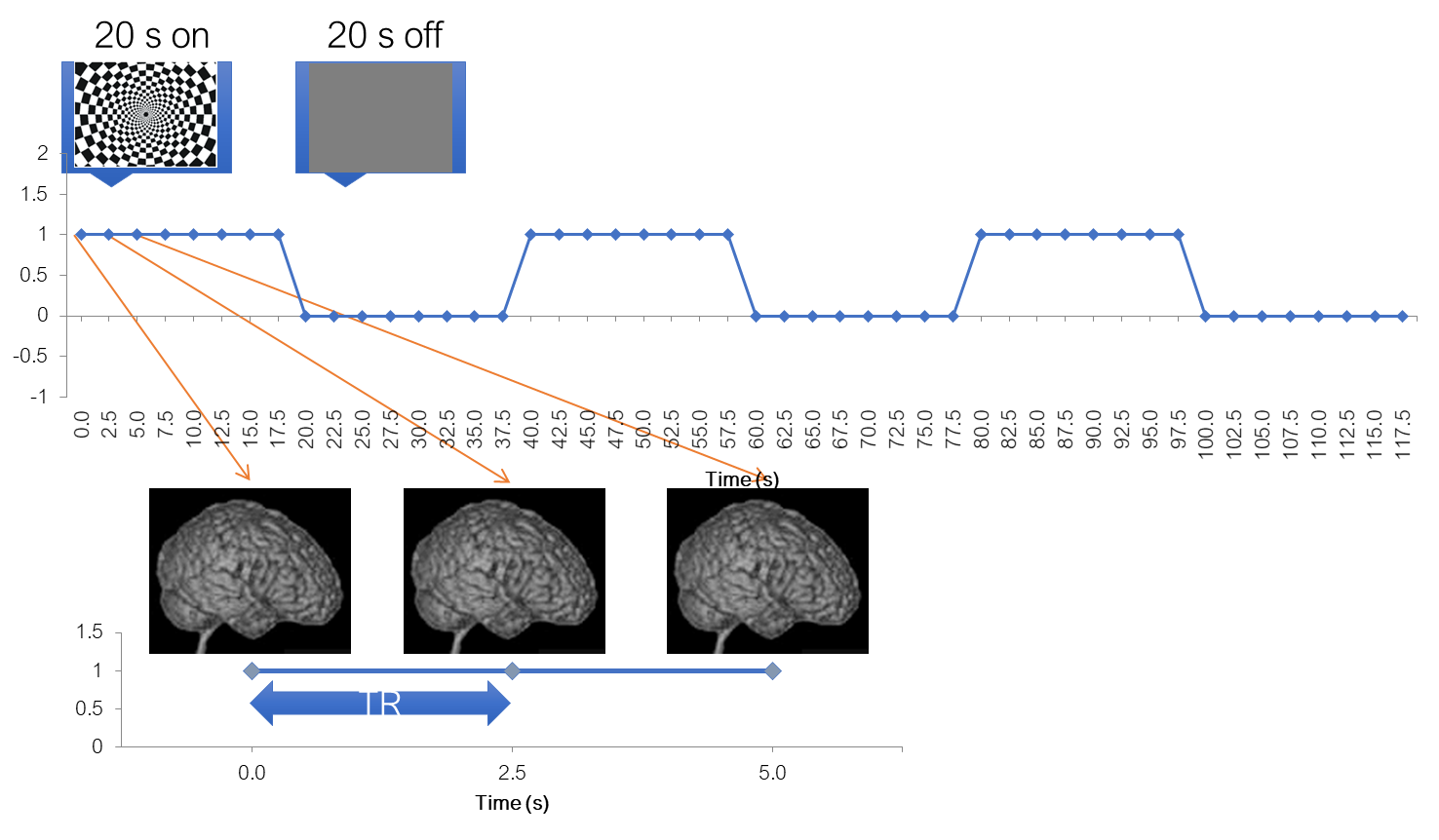
Design: Blocked or event-related
Terminology reminder
Trial
Continuous presentation of 1 experimental condition, usually 1-20 seconds
Run
Block of trials separated by interruption of a scanner acquisition, usually 5-10 minutes
Session
Block of runs, separated by subject going out of the scanner and going in again, usually at least one day